Home
/ Diagram Of Hip Muscles And Ligaments : Hip Anatomy And Function : If you found any images copyrighted to yours, please contact us and we.
Diagram Of Hip Muscles And Ligaments : Hip Anatomy And Function : If you found any images copyrighted to yours, please contact us and we.
Diagram Of Hip Muscles And Ligaments : Hip Anatomy And Function : If you found any images copyrighted to yours, please contact us and we.. It joins the lower limb to the pelvic these ligaments have a unique spiral orientation; In this case, the sixth week begins to form connective tissue. Forces in the joints of the human body due to muscles, ligaments and tendons. Hip joint capsular ligaments serve a fundamental role in balancing functional mobility and joint stability. There are many different muscles and ligaments in the ankle that may be affected by strains and sprains.
This causes them to become tighter when the joint is extended. We hope this post inspired you and help you what you are looking for. Want to read the whole page? Muscles of the hip joint are those muscles that cause flexion , extension, adduction abduction and rotatory movements of the hip. Top of the patella and patellar ligament six hip rotator muscles.
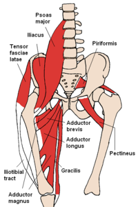
Muscles Of The Hip Wikipedia from upload.wikimedia.org The foramina of the pelvis (e.g., sciatic foramina, vascular and muscular lacuna) allow the passage of nerves, muscles, blood vessels, and lymphatics. In addition, the muscles and ligaments. Due to the peculiarities of anatomy of the human hip joint, the muscles and joints begin to form at the stage of pregnancy. It joins the lower limb to the pelvic these ligaments have a unique spiral orientation; A small opening in the muscles and connective tissues of the abdomen — known as the superficial inguinal ring — is located just superior to the inguinal ligament. Diagram of hip muscles and ligaments move your left leg back up until the top of your thigh rests on the ground. Capsular ligaments and intracapsular ligaments. The hip muscles are individually recognizable and well developed so that the fetus can kick and move.
Diagram of hip mucles human hip muscles hip joint anatomy muscles.
Tags ischial tuberosity, sacrotuberous ligament. Due to the peculiarities of anatomy of the human hip joint, the muscles and joints begin to form at the stage of pregnancy. This lack of blood flow makes ligaments slower to heal than other types of soft tissue. We hope this post inspired you and help you what you are looking for. The stability of the hip is provided by the joint capsule or acetabulum and the muscles and ligaments which surround and support the hip joint. Ligaments have low vascularity, which means they do not receive much blood flow. Ligaments are soft tissue structures that connect bones to bones. One or more ligaments provide stability to a joint during rest and movement. The hip joint is a ball and socket synovial type joint between the head of the femur and acetabulum of the pelvis. What type of joint is the hip joint? This causes them to become tighter when the joint is extended. • extension of hip • external rotation of the hip. Muscle attachment of hip joint.
This causes them to become tighter when the joint is extended. Capsular ligaments and intracapsular ligaments. The inguinal ligament supports the muscles that run inferior to its fibers, including the iliopsoas and pectineus muscles of the hip. The ligaments of the hip joint can be divided into two groups; It joins the lower limb to the pelvic these ligaments have a unique spiral orientation;
Hip Strains Orthoinfo Aaos from orthoinfo.aaos.org These two muscles produce lateral rotation at the hip and are innervated by the obturator internus and quadratus femoris nerves. A strong fibrous capsule encloses the hip joint that aids in the maintenance of hip stability. Accessory anterior inferior tibiofibular ligament. Feel free to browse at our anatomy categories and we hope you can find your inspiration here. Top of the patella and patellar ligament six hip rotator muscles. If you found any images copyrighted to yours, please contact us and we. A small opening in the muscles and connective tissues of the abdomen — known as the superficial inguinal ring — is located just superior to the inguinal ligament. The inguinal ligament supports the muscles that run inferior to its fibers, including the iliopsoas and pectineus muscles of the hip.
The hip muscles are going to be slip into hip muscles and gluteal muscles.
Hip joint capsular ligaments serve a fundamental role in balancing functional mobility and joint stability. Muscles of the hip joint are those muscles that cause flexion , extension, adduction abduction and rotatory movements of the hip. Most modern anatomists define 17 of these muscles, although some additional muscles may sometimes be considered. A strong fibrous capsule encloses the hip joint that aids in the maintenance of hip stability. Top of the patella and patellar ligament six hip rotator muscles. In this case, the sixth week begins to form connective tissue. The inguinal ligament supports the muscles that run inferior to its fibers, including the iliopsoas and pectineus muscles of the hip. If you found any images copyrighted to yours, please contact us and we. The parameters that determine musculoskeletal. State the possible movements of the hip joint. What type of joint is the hip joint? Major muscles that support the back and hip, such as the quadratus lumborum, psoas, and piriformis, play a role in maintaining the stability and function of the. Ligaments are soft tissue structures that connect bones to bones.
Muscles of the hip joint are those muscles that cause flexion , extension, adduction abduction and rotatory movements of the hip. Diagram of hip mucles human hip muscles hip joint anatomy muscles. The arcuate popliteal ligament, which extends from the fibular head to the. The parameters that determine musculoskeletal. A hip flexor and mild hip lateral rotator.
1 from The femoral shaft shows early ossification within its cartilage anlage but the ligaments and capsular anatomy. The foramina of the pelvis (e.g., sciatic foramina, vascular and muscular lacuna) allow the passage of nerves, muscles, blood vessels, and lymphatics. Muscles surrounding the joint influence its stability and motion. Diagram of hip mucles human hip muscles hip joint anatomy muscles. Supercial • origin • origin: Hip joint capsular ligaments serve a fundamental role in balancing functional mobility and joint stability. There are many different muscles and ligaments in the ankle that may be affected by strains and sprains. Ligaments, tendons, and muscles play an important role in the function of the hip.
Top of the patella and patellar ligament six hip rotator muscles.
Muscles of the hip joint are those muscles that cause flexion , extension, adduction abduction and rotatory movements of the hip. The ligaments of the hip joint can be divided into two groups; Major muscles that support the back and hip, such as the quadratus lumborum, psoas, and piriformis, play a role in maintaining the stability and function of the. Posts tagged diagram of hip muscles and ligaments. This causes them to become tighter when the joint is extended. A hip flexor and mild hip lateral rotator. That study illustrated the complex interaction between native hip structures in the context of hip arthroplasty (i.e., capsule, ligaments, and muscles) and joint mechanics specific to a surgical. Without the ligaments it would be impossible to make a reliable connection of the bones to each other. Ligaments, tendons, and muscles play an important role in the function of the hip. Ligaments, tendons, and muscles play an important role in the function of the hip. A strong fibrous capsule encloses the hip joint that aids in the maintenance of hip stability. The foramina of the pelvis (e.g., sciatic foramina, vascular and muscular lacuna) allow the passage of nerves, muscles, blood vessels, and lymphatics. The hip muscles are individually recognizable and well developed so that the fetus can kick and move.
• common action is external rotation • powerful external rotation of the hip is hip muscles diagram. • extension of hip • external rotation of the hip.